This page was reviewed under our medical and editorial policy by Maurie Markman, MD, President, Medicine & Science
This page was updated on July 20, 2022.
The gallbladder is a small, pear-shaped organ located under the liver, behind the lower-right ribs. It stores bile made by the liver. Bile is a fluid that helps to digest fats found in food, and it can be released through the common bile duct into the small intestine by either the gallbladder or the liver. Because the liver also performs this function, the gallbladder can be surgically removed without affecting a person’s health.
Gallbladder cancer is rare. Approximately 12,640 people in the United States will be diagnosed with cancer of the gallbladder and large bile duct in 2026 according to the American Cancer Society. The rate of death from this type of cancer has been decreasing in recent decades. However, because the symptoms of gallbladder cancer do not usually appear until the disease has advanced,only about 1 of 5 gallbladder cancers is found in the early stages.
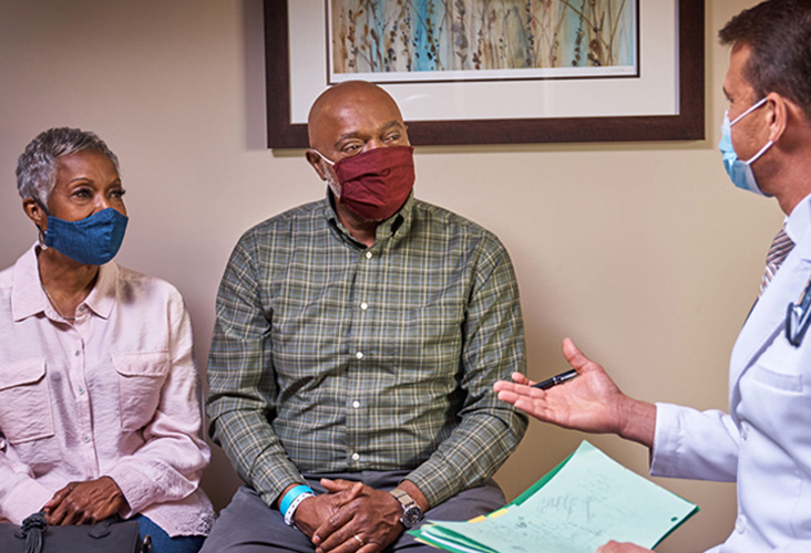
At City of Hope, we use a range of diagnostic tools and techniques to help diagnose and stage your cancer and develop a treatment plan tailored to your needs. We also offer supportive care services to help you manage the side effects of treatment, so you are better able to maintain your strength, stamina and quality of life throughout treatment.
We target gallbladder tumors with sophisticated, evidence-based treatments and technology. Your multidisciplinary team of gallbladder cancer experts will answer your questions and recommend treatment options based on your unique diagnosis and needs.
This overview will cover the basic facts about gallbladder cancer, including:
If you believe you may be experiencing symptoms of gallbladder cancer and want to schedule an appointment for diagnostic testing, or if you’re interested in a second opinion on your gallbladder cancer diagnosis and treatment plan, call us or chat online with a member of our team.
Although cancer research has not determined the causes of gallbladder cancer, certain factors may increase a person’s risk of developing the disease. Often, these factors relate to chronic inflammation of the gallbladder.
Common risk factors include:
The risk of gallbladder cancer also seems to increase in families with a history of the disease.
Gallbladder cancer occurs twice as often in women as in men. This may be related to the increased frequency of gallstones, found in up to 4 out of 5 people with gallbladder cancer. Gallstones are very common in middle-aged women, although most people with gallstones do not develop gallbladder cancer.
Gallbladder cancer develops mainly in people over the age of 65. The average age at diagnosis is 72.
Mexican Americans and Native Americans are at higher risk of gallbladder cancer, while African Americans have the lowest risk. Gallbladder cancer is less common in the United States compared with countries in Asia, Eastern Europe and South America.
People exposed to industrial chemicals, particularly those used in the rubber and textile industries, may also be at increased risk of gallbladder cancer.

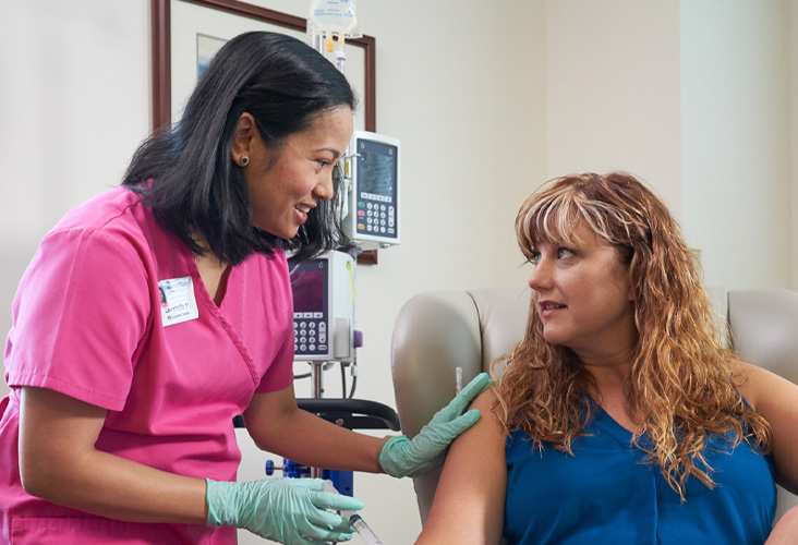
"For more than two years, I returned to City of Hope every month for five to six days at a time. I received chemotherapy intravenously and took advantage of all the supportive care services City of Hope offered."
Almost all forms of gallbladder cancer are adenocarcinomas, a type of cancer that begins in the gland-like cells that line organs of the digestive tract. Gallbladder adenocarcinomas account for 90 percent of gallbladder cancer diagnoses.
Papillary adenocarcinoma is a special subtype of adenocarcinoma that has a better prognosis compared with other types of gallbladder cancers. Its cancer cells are much less likely to spread to other parts of the body, such as nearby lymph nodes or organs.
Besides adenocarcinomas, other types of gallbladder cancer include:
Possible gallbladder cancer symptoms include:
Pathologists use these tools to diagnose gallbladder cancer:
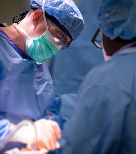
Treatment options for gallbladder cancer include:
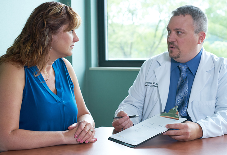
At City of Hope, we understand that gallbladder cancer and other malignancies of the gastrointestinal tract create unique challenges for patients, and that treatment options are very specific to each disease. That’s why each of our hospitals has a GI Cancer Center dedicated to diagnosing, treating and supporting the quality of life of patients with gallbladder and other GI cancers. Committed to offering state-of-the-art treatments for patients with gallbladder cancer, our multidisciplinary team of board-certified medical oncologists, surgical oncologists, radiation oncologists and supportive care clinicians work with our patients to deliver quality clinical care with a patient-centered approach. After your diagnosis, your GI Cancer Center care team will discuss your options with you and help you develop a personalized care plan tailored to your individual needs.
Because of the digestive tract’s role in processing food and waste, many patients with gastrointestinal disease have difficulty with digestive function. That’s why nutrition therapy is a key component of our GI Cancer Centers’ approach. Each center is staffed by oncology-trained dietitians who work with patients in developing a healthy, balanced and appetizing nutrition plan. If patients become malnourished, the dietitian is available to help them establish healthy lifestyle and eating habits to help improve their condition.
The GI Cancer Center teams also work closely with other supportive care clinicians to manage additional disease- and treatment-related side effects, such as nausea and vomiting. These supportive care experts may include a pain management physician, naturopathic provider, behavioral health provider and spiritual support provider.
Qualified patients may enroll in carefully selected clinical trials. Your care team will discuss whether you qualify for any of our ongoing clinical trials and, if so, help you enroll.
We understand that managing the side effects of gallbladder cancer treatment is critical to your quality of life. In addition to treating your cancer with evidence-based conventional approaches, your care team may recommend various supportive care therapies designed to help you stay strong throughout treatment. They may include: